Eyelids are what we often call eyelids. Generally speaking, they are attached to the eyeballs very naturally, forming a barrier to protect the eyeballs. There are two types of abnormal eyelids: eyelid inversion and eyelid outversion, which is also o...
Eyelids are what we often call eyelids. Generally speaking, they are attached to the eyeballs very naturally, forming a barrier to protect the eyeballs. There are two types of abnormal eyelids: eyelid inversion and eyelid outversion, which is also one of the common eye diseases in cats.
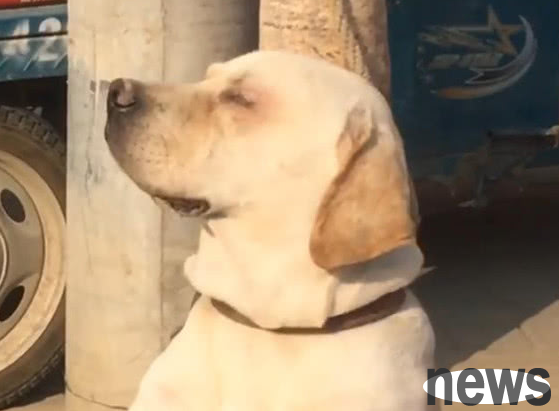
With the increase in the number of pet owners who raise cats, especially purebred cats, the number of breeds growing very rapidly. However, due to the hereditary diseases of certain breeds, many cats have eye diseases, especially many flat-faced cats often have eyelid inversion.
Once the owner is not treated in time, it will cause corneal damage. In the mild case, it will require today's medication to recover. If it is delayed a little, it will cause serious corneal ulcers, and the treatment time will last about 2 weeks. In the worse case, corneal necrosis will occur due to chronic stimulation, which is what we often call corneal rot bone.
The symptoms of eyelid inversion refer to the eyelid edge turning toward the eyeball surface, showing a series of eye disorders caused by eyelash irritation of the cornea. Most cases are caused by infection during breastfeeding, especially when the eyes are opened around 7 days, causing conjunctivitis, conjunctiva damage, and conjunctiva contraction. Clinically, it can be seen that shy tears, increase secretions, new blood vessels on the cornea, and turbid corneals. Long-term illness can cause corneal ulcers and perforations, loss of vision or loss.
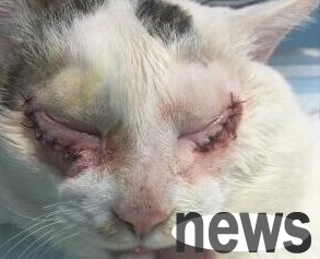
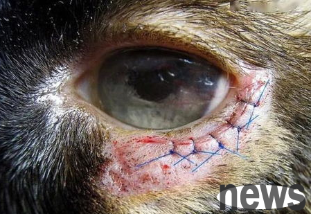
For the treatment of eyelid inversion, surgical sutures and symptomatic treatment are commonly used. For cats with mild inversion, temporary slits the skin of the eyelids and eliminates eyelash irritation to the eyeballs. For older cats, surgical correction can be performed if the valgus is severe. At 2 to 3 mm from the eye cleft, a slew-shaped skin strip is removed, the length is equal to the risk of the circumcision, and the width is to correct the circumcision. Then, the two sides of the incision are paired and three-stitch nodules are sutured. When using it, combine chloramphenicol eye drops and cortisone eye drops to alternately point the eyes.